Bioanalytics - Chemical
(M1330-06-35) In Vitro Irritability, Toxicity, and Anti-Inflammatory Activity of Rosehip Oil Emulsion
- LS
Livia Cristina Sa Barreto, Ph.D.
Professor
University of Brasilia
Brasilia, Distrito Federal, Brazil 
Joyce Santos, PhD (she/her/hers)
Nurse
Ferderal University of UFABC
Brasília, Distrito Federal, Brazil- VS
Vânia Silva, Ph.D.
Professor
Universidade Federal de Sao Paulo
São Paulo, Sao Paulo, Brazil - DO
Daniela Orsi, Ph.D.
Professor
University of Brasilia
Brasilia, Distrito Federal, Brazil - IS
Izabel Silva, Ph.D.
Professor
University of Brasilia
Brasilia, Distrito Federal, Brazil
Presenting Author(s)
Main Author(s)
Co-Author(s)
Methods:
Three different lots of emulsions containing 30% (w/w) of rosehip oil were obtained by phase inversion method using tapioca starch as a viscosity agent. An accelerated stability study was performed at 40ºC and 75% relative humidity for 90 days. The evaluation of the irritability was carried out using the chicken chorioallantoic membrane model (CAM). This test used fertilized eggs from hens incubated for 10d at 38 ± 0.5ºC and 70% relative humidity. Pure oil, oil-free emulsion, and emulsion containing 30% rosehip oil diluted in double-distilled water in a 1:1 ratio were compared with the negative control of 0.9% saline solution. The upper third of the eggshell was opened, maintaining the integrity of the embryo, until the CAM was exposed, in which 500μL of the test sample was applied. The cell viability was accessed in quintuplicate by the MTT (3-(4,5-dimethylthiazol-2-yl)-2,5-diphenyltetrazolium bromide) assay using human HaCat keratinocytes. The quantity of viable cells was determined at the wavelength of 570nm. The inhibitory concentration (IC50) was calculated using the response-sigmoid dose curve. The in vitro anti-inflammatory profile of the emulsions was investigated by the ELISA method by measurements of IL6, IL10, and TNFα cytokines. Aliquots of culture medium containing 3x10³ cells/well were seeded in 96-well plates (TPP) and incubated for 24h at 37 ºC and 5% CO2.
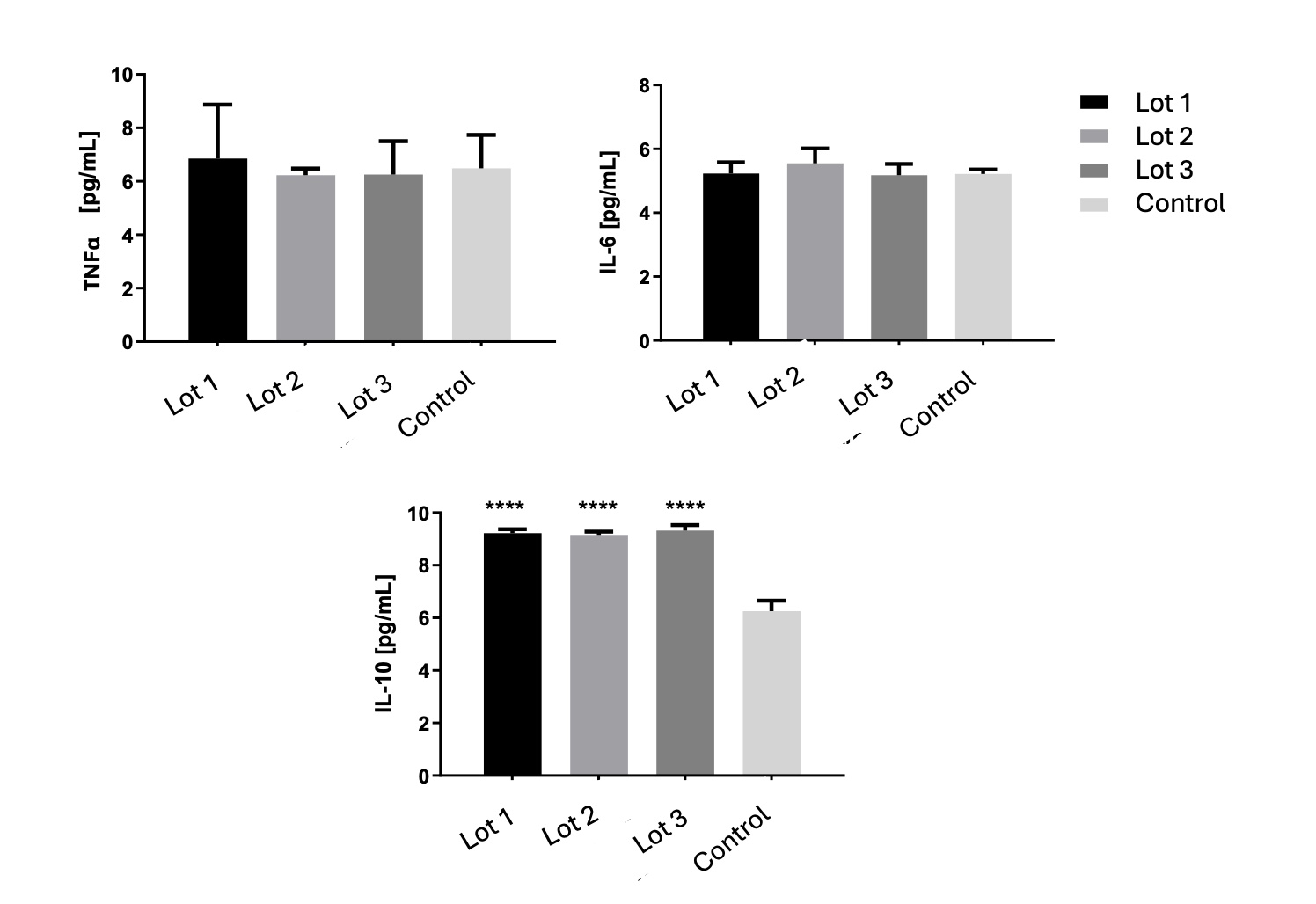
Results: The emulsions were characterized by a yellowish, consistent, opaque cream with a distinctive scent of rosehip oil. The emulsions proved to be stable from a physical-chemical point of view throughout the entire period of the accelerated stability study. Indeed, no phase separation, flocculation, or creaming occurred. The emulsions did not cause hyperemia, hemorrhage, or clotting/opacity in the chorioallantois membrane (Het-Cam test score = 0), indicating that the emulsion is no irritant. The HaCat keratinocytes did not exhibit any cytotoxicity in the cellular viability test. The emulsion needed to kill 50% of the keratinocytes was identical to that used in the control group. To be considered safe for humans, new formulas must have an inhibitory concentration above 1000 g/mL, according to the National Cancer Institute (NCI). The result found here was about four times above this limit. Regarding the anti-inflammatory activity, the emulsions significantly increased the level of IL10 compared to the negative control (Fig 1). Likewise, no alteration in the levels of TNFα and IL-6 was observed, when compared to the negative control. These results indicate that the emulsion did not stimulate the production of inflammatory cytokines, but it was able to increase IL10 release. In fact, IL10 is responsible for earlier wound healing due to its capacity to inhibit the release of inflammatory factors and activate the proliferative function of macrophages [3]. The use of rosehip oil to enhance wound healing has been frequently reported, and the findings presented here support this evidence. This effect could probably be attributed to the tocopherols present in the oil [2].
Conclusion: The right combination of emulsifiers and stabilizers, especially the use of tapioca starch as a viscosity agent, stabilized the emulsion containing a high level of rosehip oil. Despite the high amount of oil, the emulsion showed no signs of irritation or toxic behavior, suggesting its safety for human use. In addition, the emulsion's anti-inflammatory properties make it a promising vehicle for topical pharmaceutical applications such as in complex and chronic wounds.
References: 1. GONZÁLEZ-MAÑÁN, D.; ESPESSAILLES, A. D.; DOSSI, C. G.; et al. Rosa mosqueta oil prevents oxidative stress and inflammation through the upregulation of PPAR-α and NRF2 in C57BL/6J mice fed a high-fat diet. Journal of Nutrition, v. 147, n. 4, p. 579–588, 2017.
2. TAPIA, G.; SILVA, D.; ROMERO, N.; et al. Role of dietary α- and γ-tocopherol from Rosa mosqueta oil in the prevention of alterations induced by high-fat diet in a murine model. Nutrition, v. 53, p. 1–8, 2018.
3. KING, A.; BALAJI, S.; LE, L. D.; CROMBLEHOLME, T. M. Regenerative wound healing: the role of Interleukin-10. Advanves in wound care, v. 3, n. 4, p. 315–323, 2014.
Acknowledgements: The authors declare that they have no conflict of interest. The authors acknowledge the Federal District Research Support Foundation (FAP-DF, Brazil, grant n. 0019300000229/202121).
Figure 1. Cytokines levels in HaCat keratinocytes cell line supernatant. **** = P<0.0001.
