
Formulation and Delivery - Chemical

Ramkrishna Sen, PhD
Postdoctoral Fellow
University of Iowa
Iowa City, Iowa, United States
Ramkrishna Sen, PhD
Postdoctoral Fellow
University of Iowa
Iowa City, Iowa, United States
Leela Sai Lokesh Janardhanam, Ph.D.
Postdoctoral Fellow
University of Iowa
Iowa City, Iowa, United States
Mohammad Al-Natour, Ph.D.
Postdoctoral Fellow
University of Iowa
Iowa City, Iowa, United States
Md Meraj Anjum, Ph.D.
Postdoctoral Fellow
University of Iowa
Iowa City, Iowa, United States
Sean Geary, Ph.D.
Assistant Research Scientist
University of Iowa
Iowa City, Iowa, United States

Aliasger K. Salem, Ph.D.
Associate Vice President for Research
University of Iowa
Iowa City, Iowa, United States
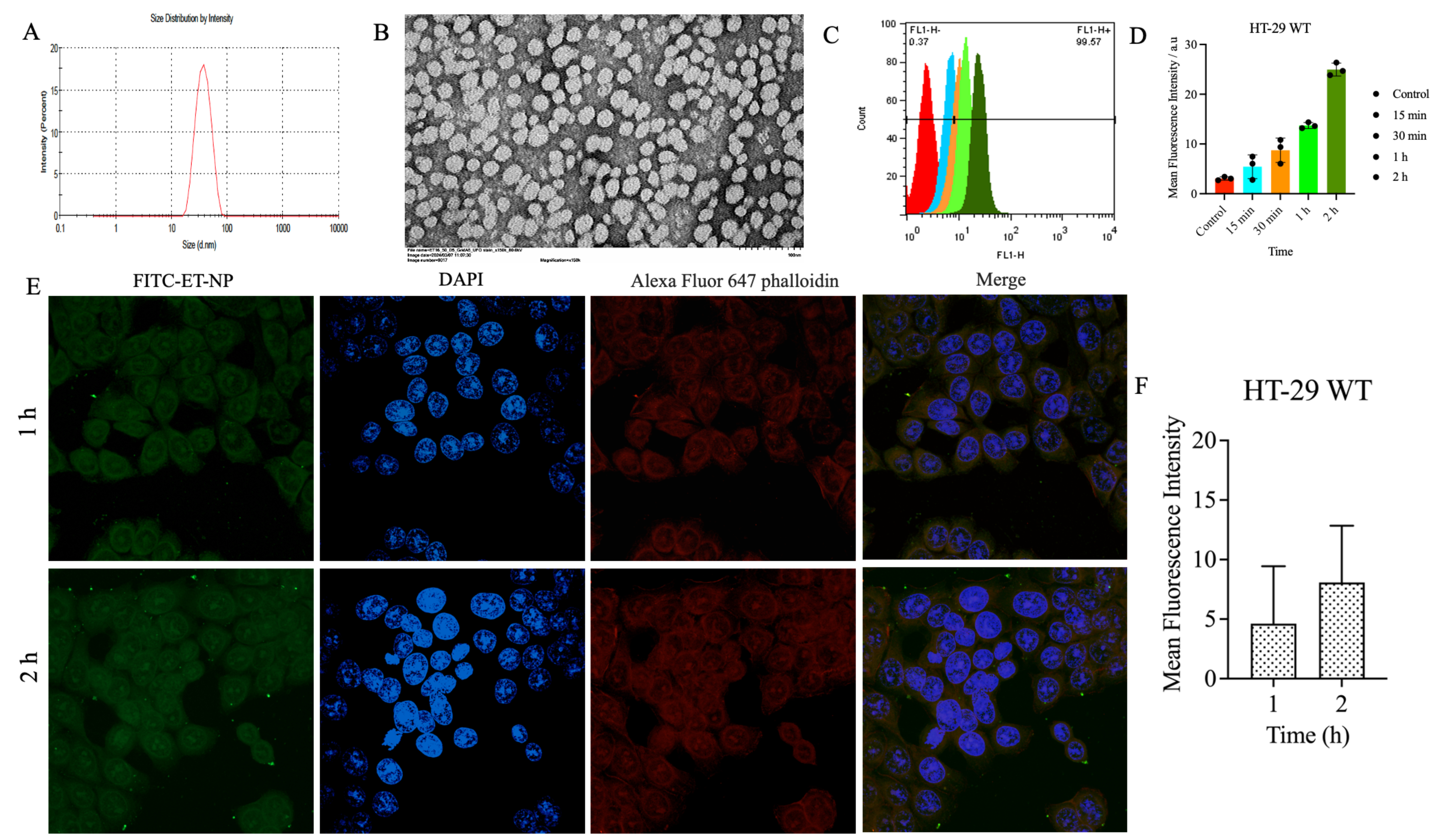 Figure 1: Physicochemical characterization and cellular uptake of experimental ET-coloaded NPs (ET-NPs). Representative figures of: (A) particle size, (B) TEM images of ET-NPs (Magnification 150X, scale bar = 100 nm). (C) Flow cytometric distribution profiles of HT-29 cells after incubation with FITC-loaded ET-NPs over time (D) Representative bar diagram depicting mean fluorescence intensity versus time for HT-29 WT after incubation with FITC-loaded ET-NPs over time. Confocal microscopic images illustrating the cellular uptake of NPs by (E) HT-29 WT cells at 1h and 2h. (F) Bar diagrams depicting the FITC mean fluorescence intensity values at different treatment durations (1h and 2h). Green color corresponds to FITC-labeled experimental NPs, red color represents F-actin, and blue color indicates the nucleus stained with DAPI. Scale bars denote 10 μm.
.jpg) Figure 2: In vivo antitumor efficacy of different treatment regimens. (A and B) Mean tumor volume recorded during the treatment period for HT-29 colon cancer xenograft mice model. (C) % reduction in tumor volume comparing the tumor volume on day 1 and day 13 of treatment. The negative values indicate that tumor volume increased compared to the initial volume. (D) Body weight recorded during the treatment period for the HT-29 colon cancer xenograft mice model. Data are plotted as mean ± SD (n = 3). One-way ANOVA and unpaired t-test were used for statistical analysis. *, < 0.0186: **, < 0.0070: ***, < 0.0006: non-significant (ns).
Figure 2: In vivo antitumor efficacy of different treatment regimens. (A and B) Mean tumor volume recorded during the treatment period for HT-29 colon cancer xenograft mice model. (C) % reduction in tumor volume comparing the tumor volume on day 1 and day 13 of treatment. The negative values indicate that tumor volume increased compared to the initial volume. (D) Body weight recorded during the treatment period for the HT-29 colon cancer xenograft mice model. Data are plotted as mean ± SD (n = 3). One-way ANOVA and unpaired t-test were used for statistical analysis. *, < 0.0186: **, < 0.0070: ***, < 0.0006: non-significant (ns). .jpg) Tables: 1 Physicochemical Evaluation of Formulations: Data from the three independent experiments. E = encorafenib, T = trametinib
Tables: 1 Physicochemical Evaluation of Formulations: Data from the three independent experiments. E = encorafenib, T = trametinib