
Formulation and Delivery - Chemical
.jpg "Hytham H. Gadalla, MS (he/him/his) photo")
Hytham H. Gadalla, MS (he/him/his)
Graduate student
Purdue University
West Lafayette, Indiana, United States
Hytham H. Gadalla, MS (he/him/his)
Graduate student
Purdue University
West Lafayette, Indiana, United States
Marwa G. Elnaggar, Ph.D. (she/her/hers)
Postdoc Research Associate
Purdue University
West Lafayette, Indiana, United States
 photo")
Fanfei Meng, Ph.D. (he/him/his)
Assistant Professor
University of Massachusetts
Lowell, Massachusetts, United States

Yoon Yeo, Ph.D.
Associate Department Head, Industrial and Molecular Pharmaceutics
Purdue University
West Lafayette, Indiana, United States
.jpg) Fig. 1. Preparation and in vitro characterization of Nanosac. (a) Schematic description of Nanosac preparation. (b) Size and surface charge by DLS. (c) Morphology by TEM. (d) Deformability by AFM. (e) Colloidal stability in different media. (f) Cell viability by MTT assay. (g) Checkpoint protein silencing efficiency. (h) PD-L1 silencing activity of lyophilized Nanosac. (i) Phagocytosis of B16F10 tumor cells by J774a.1 macrophages evaluated with confocal microscopy. (j) Redox-responsive siRNA release from Nanosac.
Fig. 1. Preparation and in vitro characterization of Nanosac. (a) Schematic description of Nanosac preparation. (b) Size and surface charge by DLS. (c) Morphology by TEM. (d) Deformability by AFM. (e) Colloidal stability in different media. (f) Cell viability by MTT assay. (g) Checkpoint protein silencing efficiency. (h) PD-L1 silencing activity of lyophilized Nanosac. (i) Phagocytosis of B16F10 tumor cells by J774a.1 macrophages evaluated with confocal microscopy. (j) Redox-responsive siRNA release from Nanosac.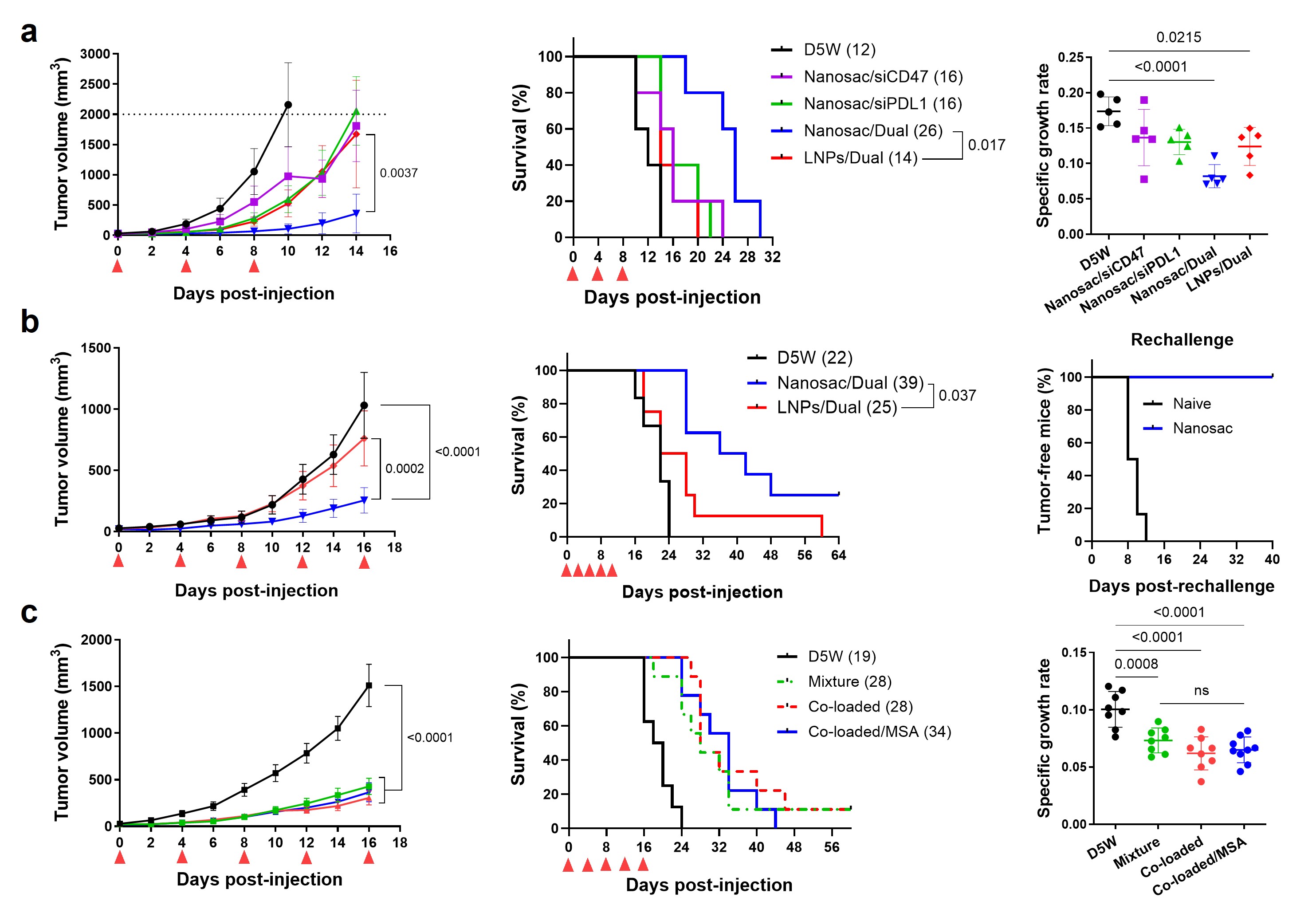 Fig. 2. In vivo antitumor efficacy of Nanosac. Antitumor efficacy in subcutaneous mouse models of (a) B16F10 melanoma or (b and c) CT26 colon carcinoma. Red arrowheads: IV dosing. Numbers in parenthesis: median survival time in days.
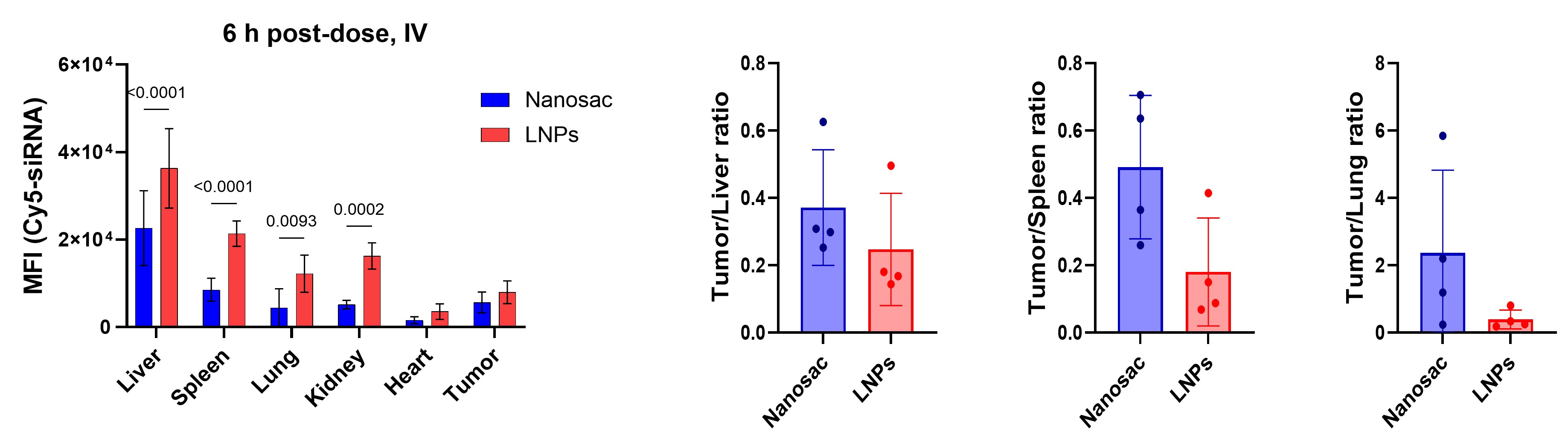 Fig. 3. Biodistribution of Cy5-siRNA loaded Nanosac or LNPs in subcutaneous CT26 tumor bearing mice 6 hours post-IV injection. Right: Mean fluorescence intensity in organ homogenates; Left: Fluorescence signal in tumors normalized to that in the MPS organs.