
Formulation and Delivery - Biomolecular
Zhongyue Yuan, MS
Graduate Student
Purdue University
West Lafayette, Indiana, United States
Zhongyue Yuan, MS
Graduate Student
Purdue University
West Lafayette, Indiana, United States
Wenzhu Qi, MS
Graduate Student
Purdue University
West Lafayette, Indiana, United States
Morgan J. Robinson, Ph.D.
Post doc
Purdue University
West Lafayette, Indiana, United States
Jean Christophe Rochet, Ph.D.
Professor of Medicinal Chemistry and Molecular Pharmacology
Purdue University
West Lafayette, Indiana, United States
Yang Yang, Ph.D.
Associate Professor of Medicinal Chemistry and Molecular Pharmacology
Purdue University
West Lafayette, Indiana, United States

Yoon Yeo, Ph.D.
Associate Department Head, Industrial and Molecular Pharmaceutics
Purdue University
West Lafayette, Indiana, United States
.jpg) Fig.1. In vitro characterization and optimization of Nanosac. (a) Z-average and zeta potential of Nanosac. (b) Cytotoxicity of Nanosac on hCMEC/d3, and (c) SH-SY5Y cells. (d) Effect of pre-albuminylation on Nanosac colloidal stability. (e) Effect of PVA on the particle size of Nanosac.
Fig.1. In vitro characterization and optimization of Nanosac. (a) Z-average and zeta potential of Nanosac. (b) Cytotoxicity of Nanosac on hCMEC/d3, and (c) SH-SY5Y cells. (d) Effect of pre-albuminylation on Nanosac colloidal stability. (e) Effect of PVA on the particle size of Nanosac. 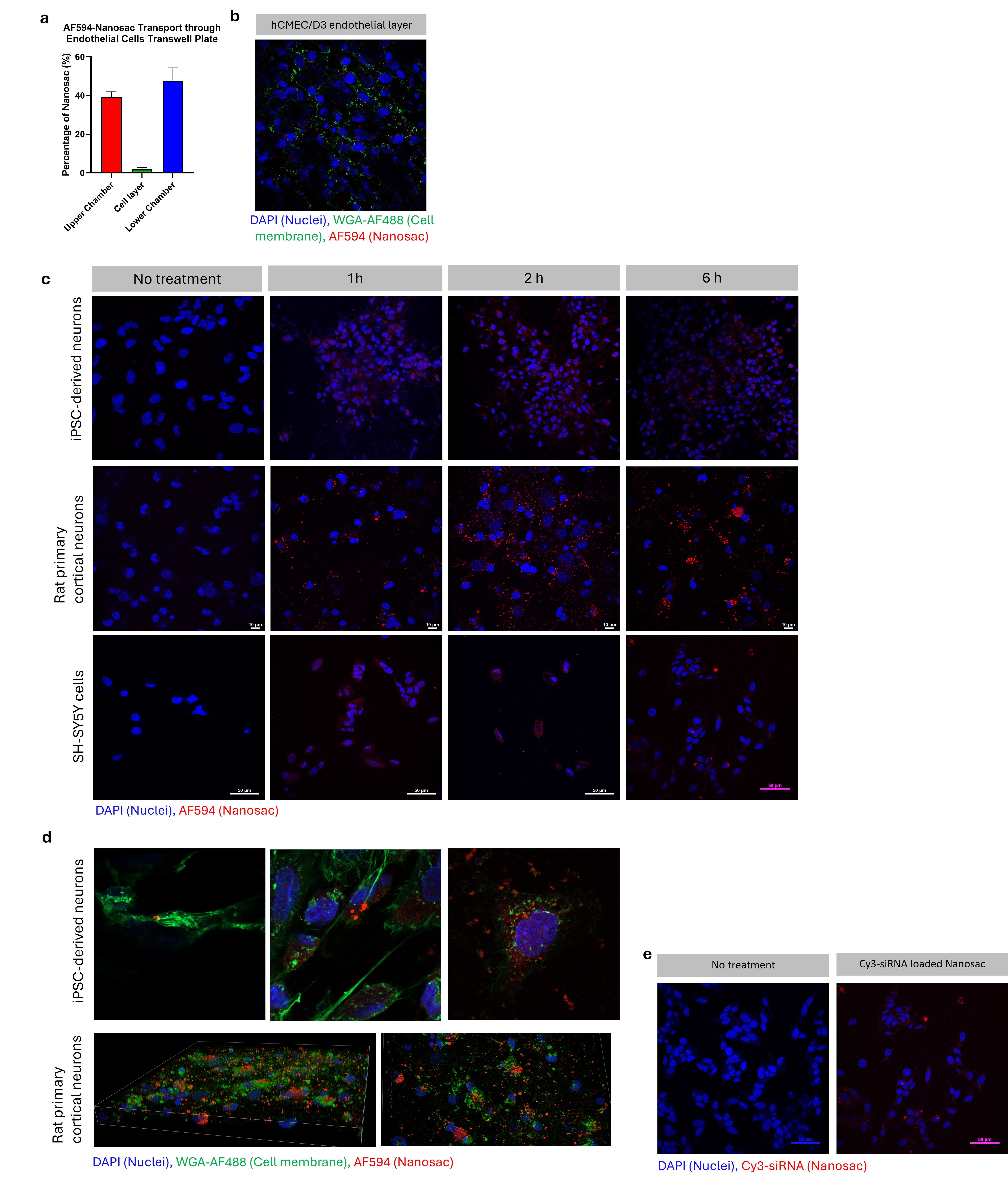 Fig. 2. Nanosac transport across the endothelial layer and cell uptake by neurons. (a) The percentage of Nanosac transport across the confluent hCMEC/d3 endothelial layer. (b) Neurons on Transwell membrane. Blue: DAPI (Nuclei); Green: Wheat Germ Agglutinin (WGA)-AF488 (Cell membrane); Red: AF594 (Nanosac) (c) Nanosac cell uptake by iPSC-derived neurons, rat primary cortical neurons, and SH-SY5Y cell in 1, 2, and 6 h. Blue: DAPI (Nuclei); Red: AF594 (Nanosac) (d) Nanosac in the cell body and projections after incubation for 6 h. Blue: DAPI (Nuclei); Green: WGA-AF488 (Cell membrane); Red: AF594 (Nanosac) (e) Cy3-siRNA delivered by Nanosac to SH-SY5Y cells after incubation for 6 h. Blue: DAPI (Nuclei); Red: Cy3-siRNA.
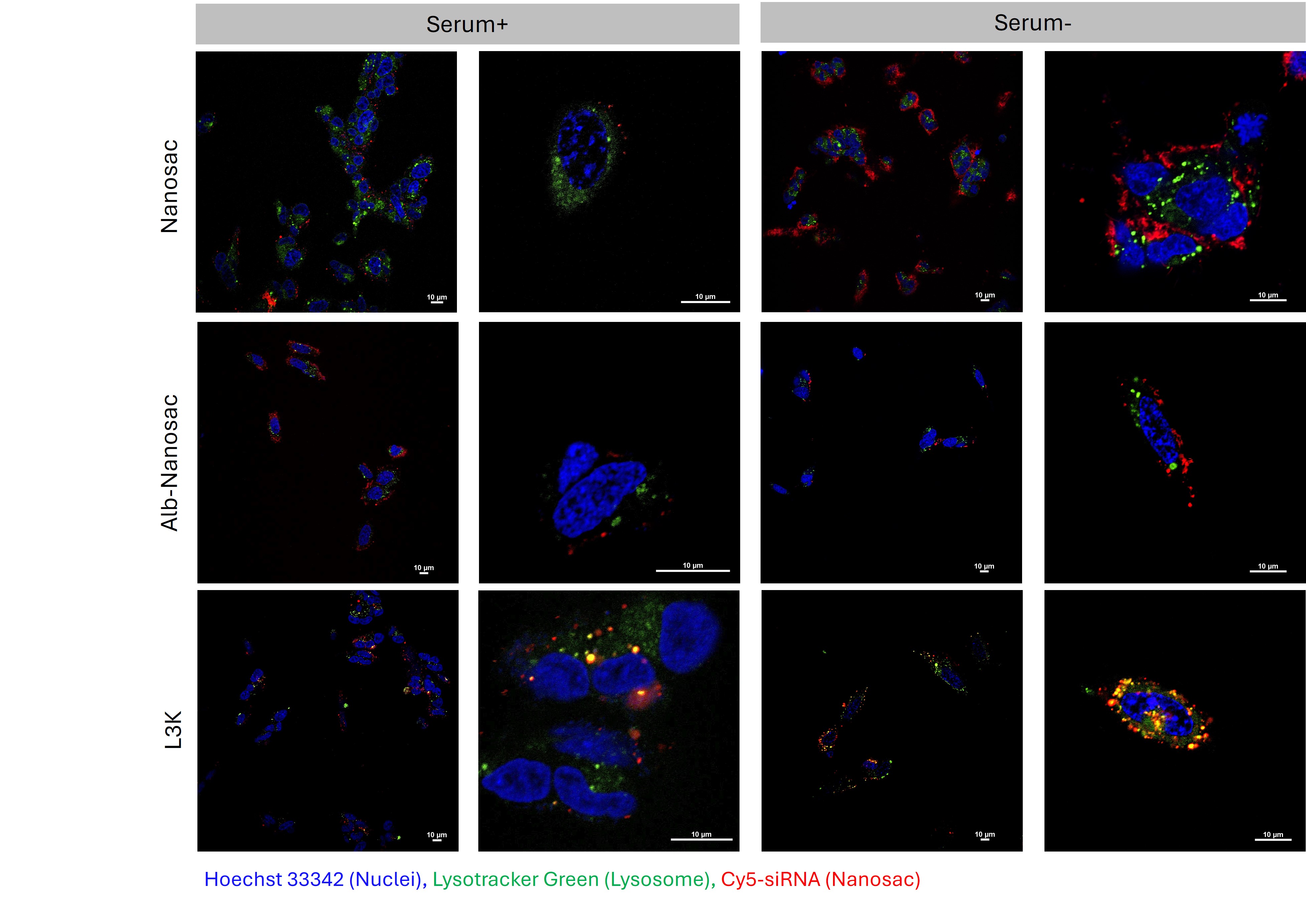 Fig. 3. Confocal microscope images locating Cy5-siRNA loaded Nanosac, Alb-Nanosac, and L3K relative to lysosomes in SH-SY5Y cells. Blue: Hoechst 33342 (nuclei); Green: Lysotracker (lysosome); red: Cy5-siRNA. Incubation time: 6 h. Scale bars: 10 μm.