
Formulation and Delivery - Biomolecular
Sara Aly Attia, BS
Ph.D. Candidate
University of Southern California
Los Angeles, California, United States
J. Andrew MacKay, Ph.D.
Professor
University of Southern California
N/A, California, United States
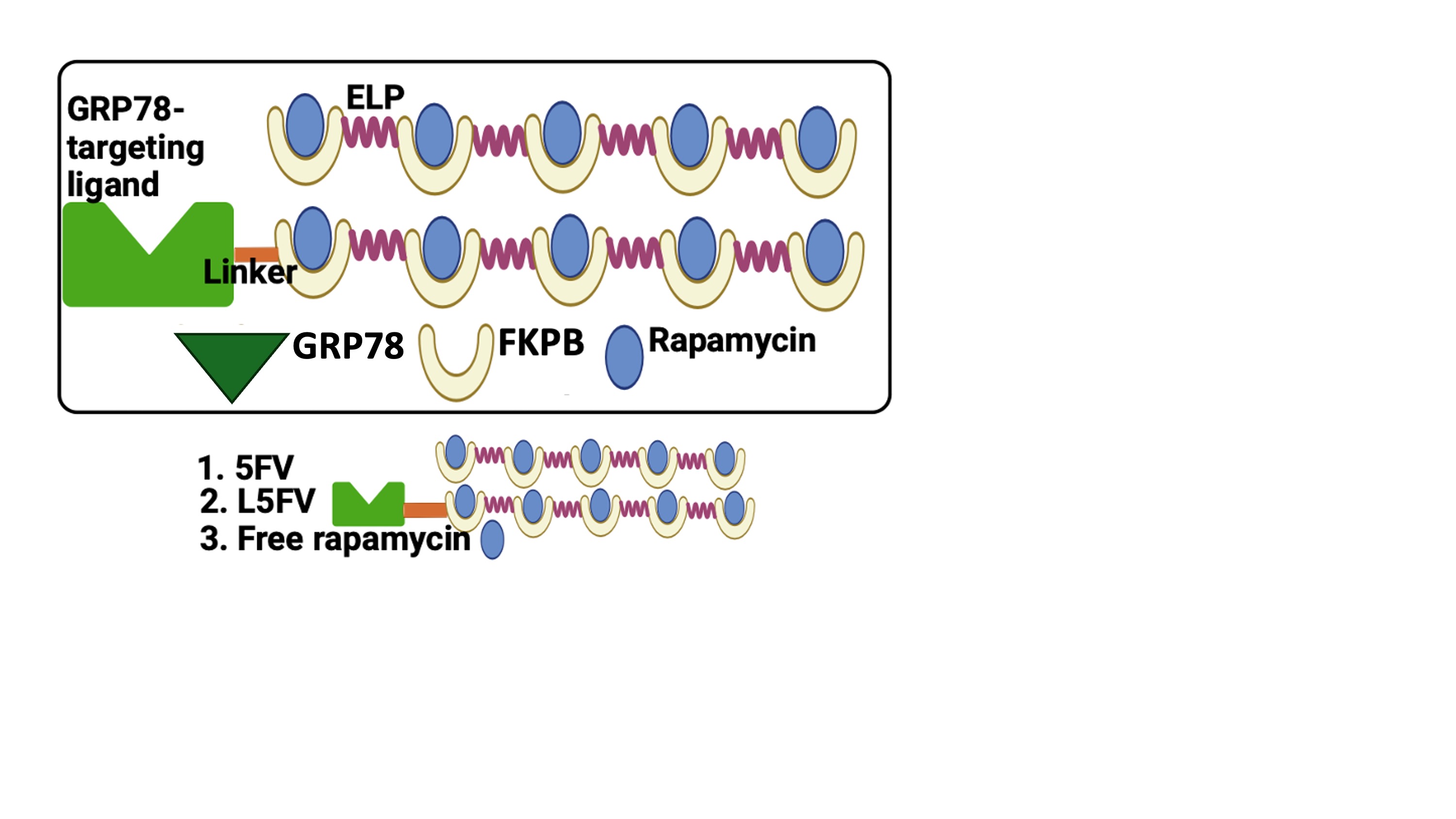 Figure 1. GRP78-targeting FKPB-ELP fusions for treatment groups employed in the study (GRP78-targeted L-5FV, Untargeted 5FV, Free Rapa).
 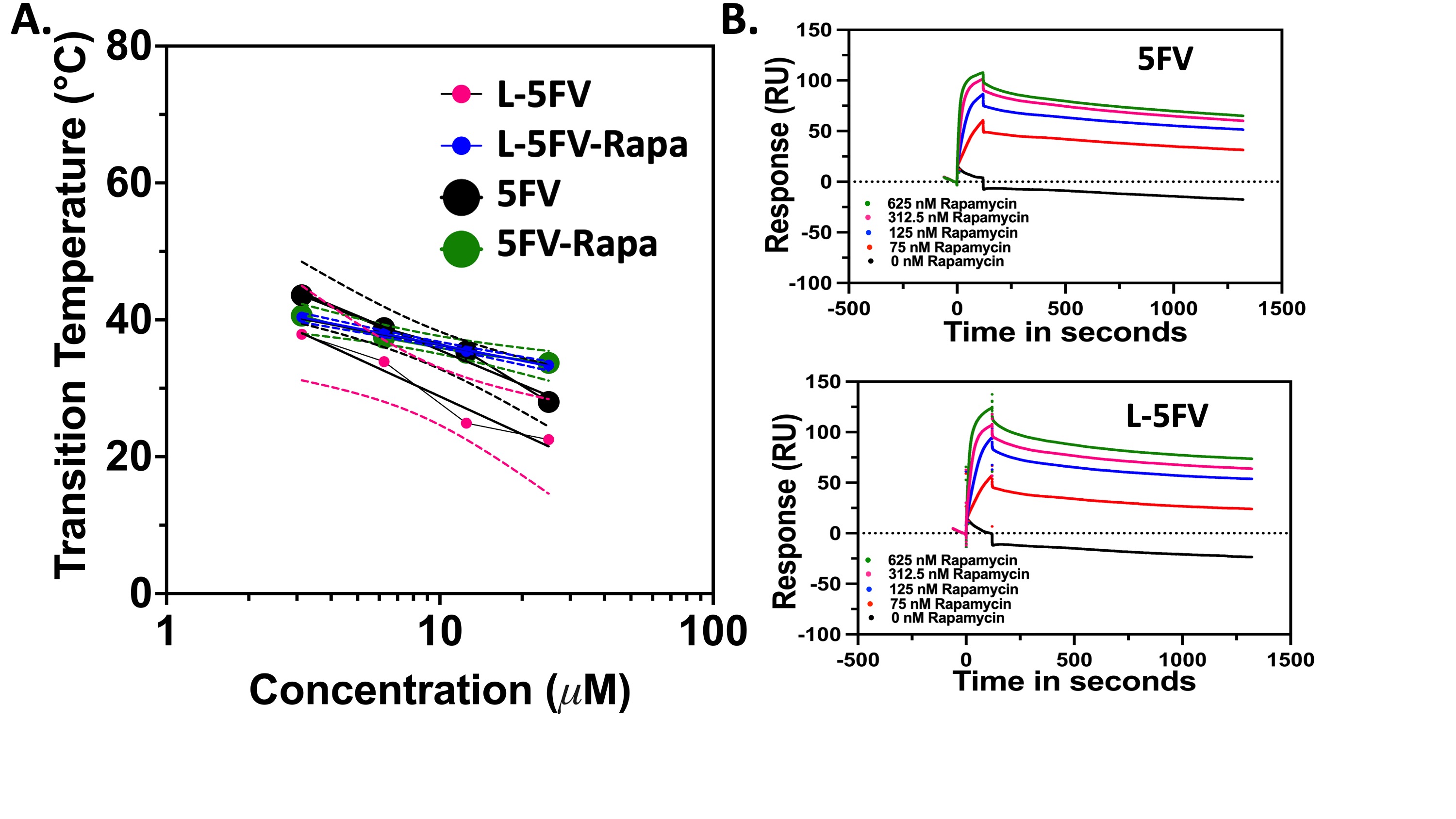 Figure 2. Rapa binds to FKBP-ELPs with and without the GRP78 targeting L-peptide. Rapa binding slightly raises the phase transition temperature of 5FV and L-5FV fusions. A) Optical-density was used to measure Tt for purified 5FV, L-5FV, 5FV-Rapa, and L-5FV-Rapa formulations (15-80 ºC) at a wavelength of 350 nm over a range of concentrations (25, 12.5, 6.25, and 3.13 µM) in PBS. The maximum first derivative was defined as Tt. As shown, Tt was log-linear with the concentration. B) SPR quantified the affinity of 5FV and L-5FV to Rapa over a range of concentrations (625, 312.5, 125, and 75 nM) below Tt. Rapa bound to 5FV and L-5FV with less than 1 nM equilibrium binding constant.
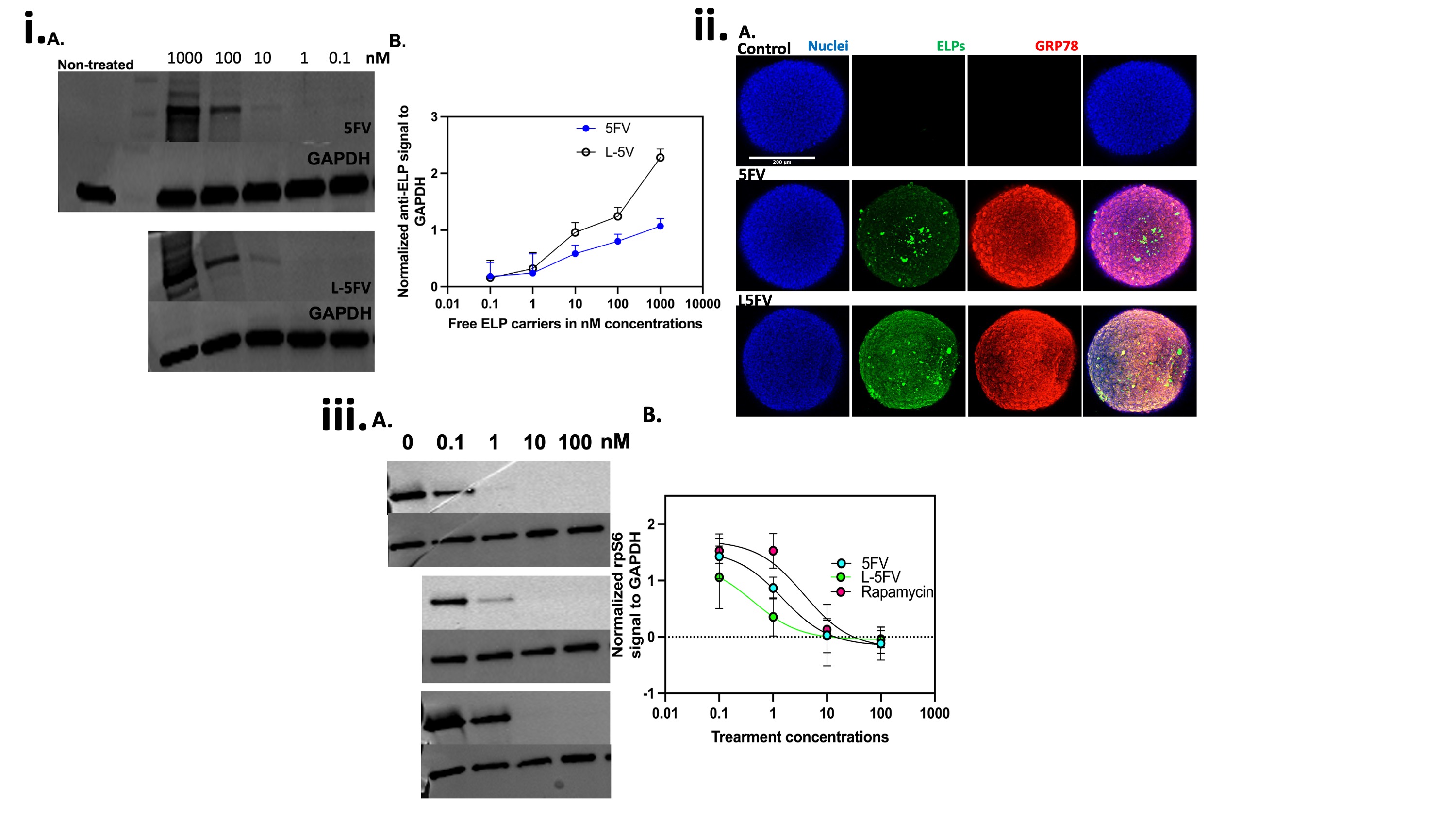 Figure 3. L-5FV/Rapa undergoes more dose-dependent cell association and inhibition of P-rpS6 than 5FV. Rapa inhibits cancer proliferation through inhibition of mTORC-1 in BT474 cells. Compared to 5FV, i) Western blotting against ELP using an anti-ELP antibody and ii) confocal microscopy of 3D spheroids stained using an anti-ELP antibody revealed more L-5FV cellular association at 10 to 1000 nM concentrations. L-5FV/Rapa had significant cellular internalization at the spheroid surface as well as the interior compared to 5FV/Rapa. iii) Demonstrating their relative delivery of Rapa, BT474 cells were incubated with 100, 10, 1, and 0.1 nM Rapa bound to 5FV, L-5FV or free Rapa. P-rpS6 signal by WB was normalized to GAPDH, which revealed the most potent suppression mTORC-1 for GRP-78 targeted L-5FV (**P = 0.001 AND *P = 0.04), relative to 5FV-Rapa AND free Rapa groups, respectively.