
Formulation and Delivery - Chemical
Sumedha Kapre, MS
Graduate Student
Texas A&M University
KINGSVILLE, Texas, United States
Sumedha Kapre, MS
Graduate Student
Texas A&M University
KINGSVILLE, Texas, United States
Sushesh Srivatsa Palakurthi, MS
Graduate Student
Texas A&M University
Kingsville, Texas, United States
Mrudul Velhal, MS
Graduate Student
Texas A&M University
College Station, Texas, United States
Hong Liang, Ph.D.
Professor
Texas A&M University
College Station, Texas, United States
Wei Zheng, Ph.D.
Assistant Professor
Texas A&M University
Corpus Christi, Texas, United States
Srinath Palakurthi, Ph.D. (he/him/his)
Professor
Texas A&M University
Kingsville, Texas, United States
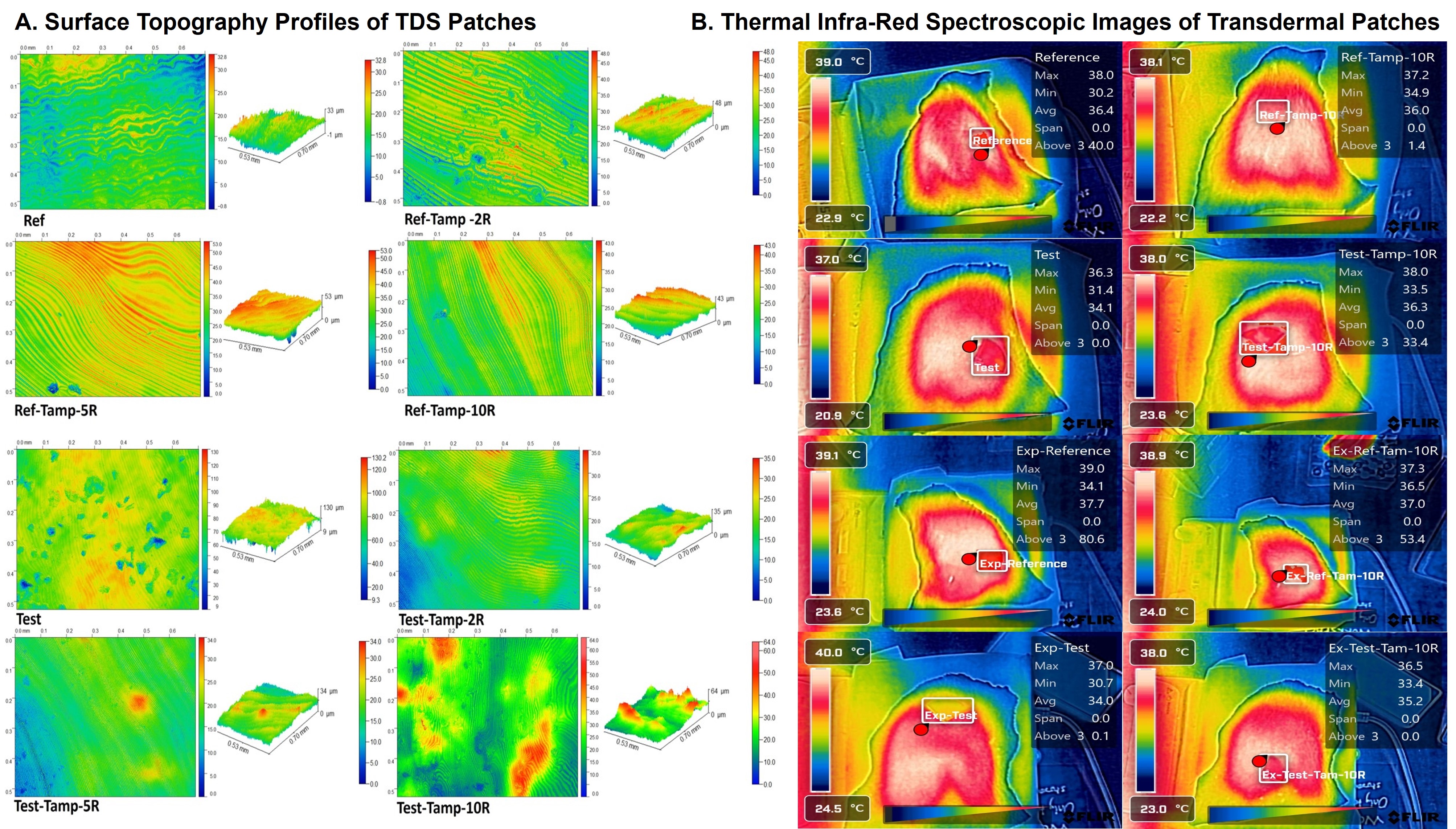 Figure 1: Surface topography, roughness profiles, and thermal infra-red spectroscopic images of transdermal patches. (A). Surface topography, and roughness profiles of Reference and Test patches. Un-expired, 2R, 5R, and 10R tampered patches were tested (n=3; p=0.0093, 95% CI). (B). Infra-red thermal imaging was conducted for the same sets of variables. The mean difference in the surface temperature of the skin where the patch was adhered was recorded (n=3). The images were captured when the temperature was at least above 37±1 °C.
 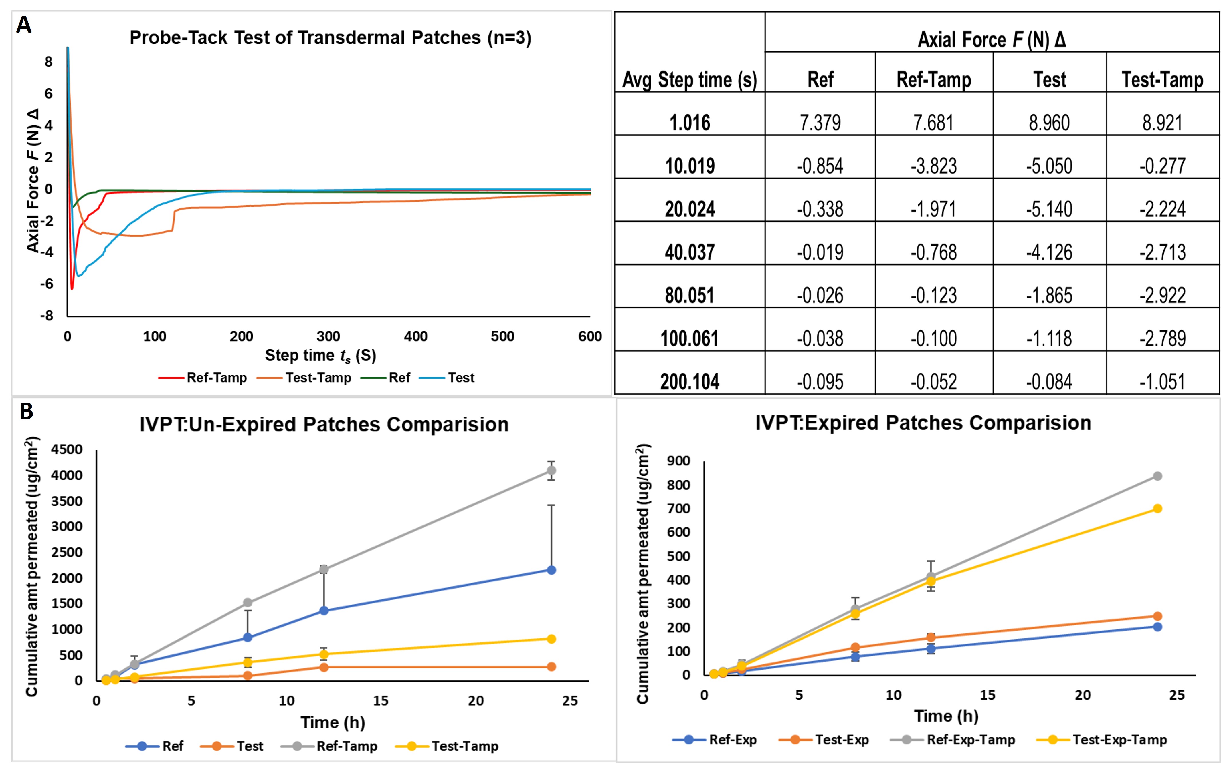 Figure 2: Adhesiveness performance evaluation and in vitro drug permeation testing of transdermal patches. (A). Adhesiveness performance was evaluated by probe-tack test using a TA instruments DHR-2 Rheometer. A 8 mm stainless steel peltier plate, with a diameter of 40 mm, loading gap of 30 mm was maintained. An axial force of 10N was applied for 30 seconds, and the experiment was conducted at 25 °C (n=3) (B). In vitro permeation testing of the reference and test transdermal patches were conducted using a vertical Franz diffusion cell of 1.77 cm2 receiver cell orifice area. Porcine ear explant was used, and the experiments were conducted at 37±0.5°C (n=3, p<0.5).