
Formulation and Delivery - Biomolecular
 photo")
Jungmi Park (she/her/hers)
Graduate Student
University of Massachusetts
Belchertown, Massachusetts, United States
Jungmi Park (she/her/hers)
Graduate Student
University of Massachusetts
Belchertown, Massachusetts, United States
Taewon Jeon, Ph.D.
Post-Doc
Massachusetts Institute of Technology
Amherst, Massachusetts, United States
Jessa Marie V.Makabenta, Ph.D.
Senior R&D Chemist
Avient
Amherst, Massachusetts, United States
Vincent M. Rotello, Ph.D.
Professor
University of Massachusetts
Amherst, Massachusetts, United States
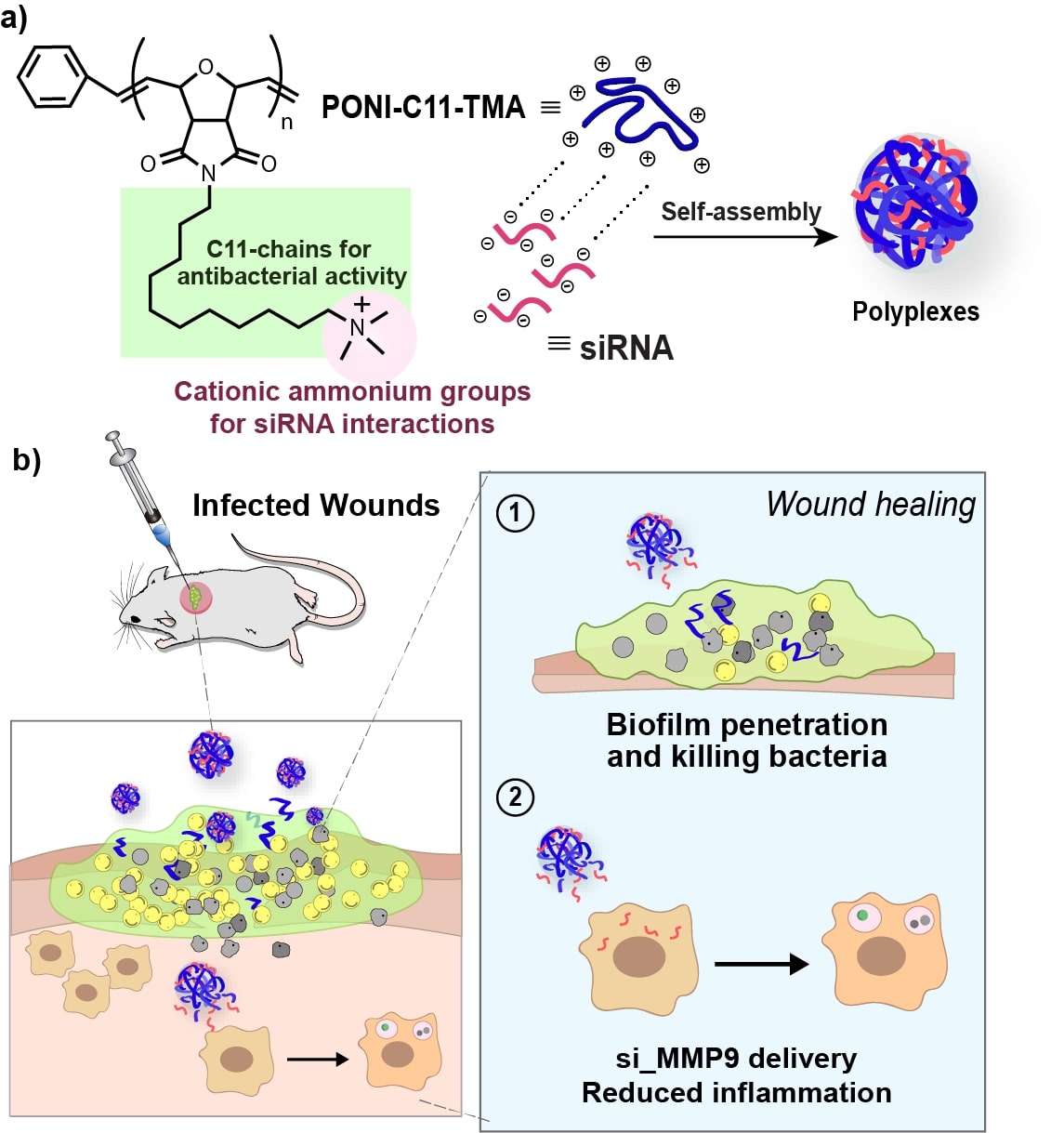 Figure 1. Schematic representation of a) engineering polyplexes via electrostatic interactions of siRNA and PONI-C11-TMA and b) in vivo treatment of polyplexes for infected wounds on mice showing efficient biofilm penetration and eradication of bacteria combined with si_MMP9 delivery strategy induced reduction in inflammation.
.jpg) Figure 3. Effects of polyplexes on methicillin-resistant S. aureus (MRSA) biofilm. a) Representative 3D views of confocal image stacks of red fluorescent protein (RFP)-expressing MRSA biofilm after 1 h incubation with Cy5.5-labelled polyplexes (Cyan). Overlay images show Cy5.5-polyplexes completely penetrate the entire biofilm, interacting with MRSA cells. Biofilm thickness is ~ 18μm. b) Screening the polymer and the polyplexes formulated with different N/P ratios via their antimicrobial activity against MRSA IDRL-6169 for biocompatibility. c) Evaluating siRNA activity through MMP9 knockdown in RAW 264.7 macrophages quantified by qRT-PCR. Error bars represent the standard deviation (SD) of three experimental replicates (Data are presented as mean ± SD, one-way ANOVA, and Tukey multiple comparisons, ****p < 0.001). and fluorescent reporter gene silencing and evaluation of siRNA activity. d) Representative confocal microscopy images of cells after treatment with PONI-C11-TMA/si_eGFP polyplexes. Cell nuclei stained with DAPI (Blue). Deliveries were performed with polyplexes formulated at N/P 40 ratio with 100 nM of siRNA. Scale bar: 50 μm.
Figure 3. Effects of polyplexes on methicillin-resistant S. aureus (MRSA) biofilm. a) Representative 3D views of confocal image stacks of red fluorescent protein (RFP)-expressing MRSA biofilm after 1 h incubation with Cy5.5-labelled polyplexes (Cyan). Overlay images show Cy5.5-polyplexes completely penetrate the entire biofilm, interacting with MRSA cells. Biofilm thickness is ~ 18μm. b) Screening the polymer and the polyplexes formulated with different N/P ratios via their antimicrobial activity against MRSA IDRL-6169 for biocompatibility. c) Evaluating siRNA activity through MMP9 knockdown in RAW 264.7 macrophages quantified by qRT-PCR. Error bars represent the standard deviation (SD) of three experimental replicates (Data are presented as mean ± SD, one-way ANOVA, and Tukey multiple comparisons, ****p < 0.001). and fluorescent reporter gene silencing and evaluation of siRNA activity. d) Representative confocal microscopy images of cells after treatment with PONI-C11-TMA/si_eGFP polyplexes. Cell nuclei stained with DAPI (Blue). Deliveries were performed with polyplexes formulated at N/P 40 ratio with 100 nM of siRNA. Scale bar: 50 μm. .jpg) In vivo therapeutic efficacy of PONI-C11-TMA/siRNA polyplexes for severe wound biofilm infections. a) Schematic representation of the murine model of wound biofilm infection. b) SEM image of mice skin sample confirming biofilm formation at the wound site. Scale bar: 5 μm. c) Bioluminescence signals from the wound area of representative mice on the different days of treatment. d) Extent of bacterial reduction relative to negative control, PBS only [bacteria reduction = (log CFU count)PBS - (log CFU count)treatment group]. Error bars represent the mean ± the standard error of the mean (SEM) (n=4, one-way ANOVA and Tukey multiple comparisons, *p < 0.05). e) In vivo treatment of PONI-C11-TMA/si_MMP9 decreased MMP9 mRNA levels quantified by qRT-PCR. Error bars represent the mean ± the standard error of the mean (SEM) (n=4, one-way ANOVA and Tukey multiple comparisons, ***p < 0.001. f) Degree of wound size reduction at the day of sacrifice relative to the first day of treatment. One-way ANOVA and Tukey multiple comparisons, *, **, *** indicate p-value < 0.05, 0.01 or 0.001, respectively.
In vivo therapeutic efficacy of PONI-C11-TMA/siRNA polyplexes for severe wound biofilm infections. a) Schematic representation of the murine model of wound biofilm infection. b) SEM image of mice skin sample confirming biofilm formation at the wound site. Scale bar: 5 μm. c) Bioluminescence signals from the wound area of representative mice on the different days of treatment. d) Extent of bacterial reduction relative to negative control, PBS only [bacteria reduction = (log CFU count)PBS - (log CFU count)treatment group]. Error bars represent the mean ± the standard error of the mean (SEM) (n=4, one-way ANOVA and Tukey multiple comparisons, *p < 0.05). e) In vivo treatment of PONI-C11-TMA/si_MMP9 decreased MMP9 mRNA levels quantified by qRT-PCR. Error bars represent the mean ± the standard error of the mean (SEM) (n=4, one-way ANOVA and Tukey multiple comparisons, ***p < 0.001. f) Degree of wound size reduction at the day of sacrifice relative to the first day of treatment. One-way ANOVA and Tukey multiple comparisons, *, **, *** indicate p-value < 0.05, 0.01 or 0.001, respectively.