
Formulation and Delivery - Biomolecular
 photo")
Joan Cheng, BS (she/her/hers)
PhD Student
University of Minnesota
Minneapolis, Minnesota, United States
Joan Cheng, BS (she/her/hers)
PhD Student
University of Minnesota
Minneapolis, Minnesota, United States
Karunya K. Kandimalla, Ph.D.
Associate dean of graduate studies, Associate Professor
University of Minnesota
Minneapolis, Minnesota, United States
.jpg) (Left) Micrographs of polarized hCMEC/D3 monolayers after clathrin or caveolin-1 knockdown and treatment with cationic bicelles. Nuclei were stained with DAPI (blue), bicelles were laced with DiI (yellow), and clathrin or caveolin-1 were tagged with Alexa Fluor 647 (red). The scale bar is 20 μm. (Right) Protein expression and bicelle uptake in clathrin or caveolin-1 knockdown hCMEC/D3 cells expressed as a percentage relative to the wild-type. Values were quantified based on mean fluorescence and normalized to mean DAPI fluorescence. Mean ± SD (n = 6).
(Left) Micrographs of polarized hCMEC/D3 monolayers after clathrin or caveolin-1 knockdown and treatment with cationic bicelles. Nuclei were stained with DAPI (blue), bicelles were laced with DiI (yellow), and clathrin or caveolin-1 were tagged with Alexa Fluor 647 (red). The scale bar is 20 μm. (Right) Protein expression and bicelle uptake in clathrin or caveolin-1 knockdown hCMEC/D3 cells expressed as a percentage relative to the wild-type. Values were quantified based on mean fluorescence and normalized to mean DAPI fluorescence. Mean ± SD (n = 6).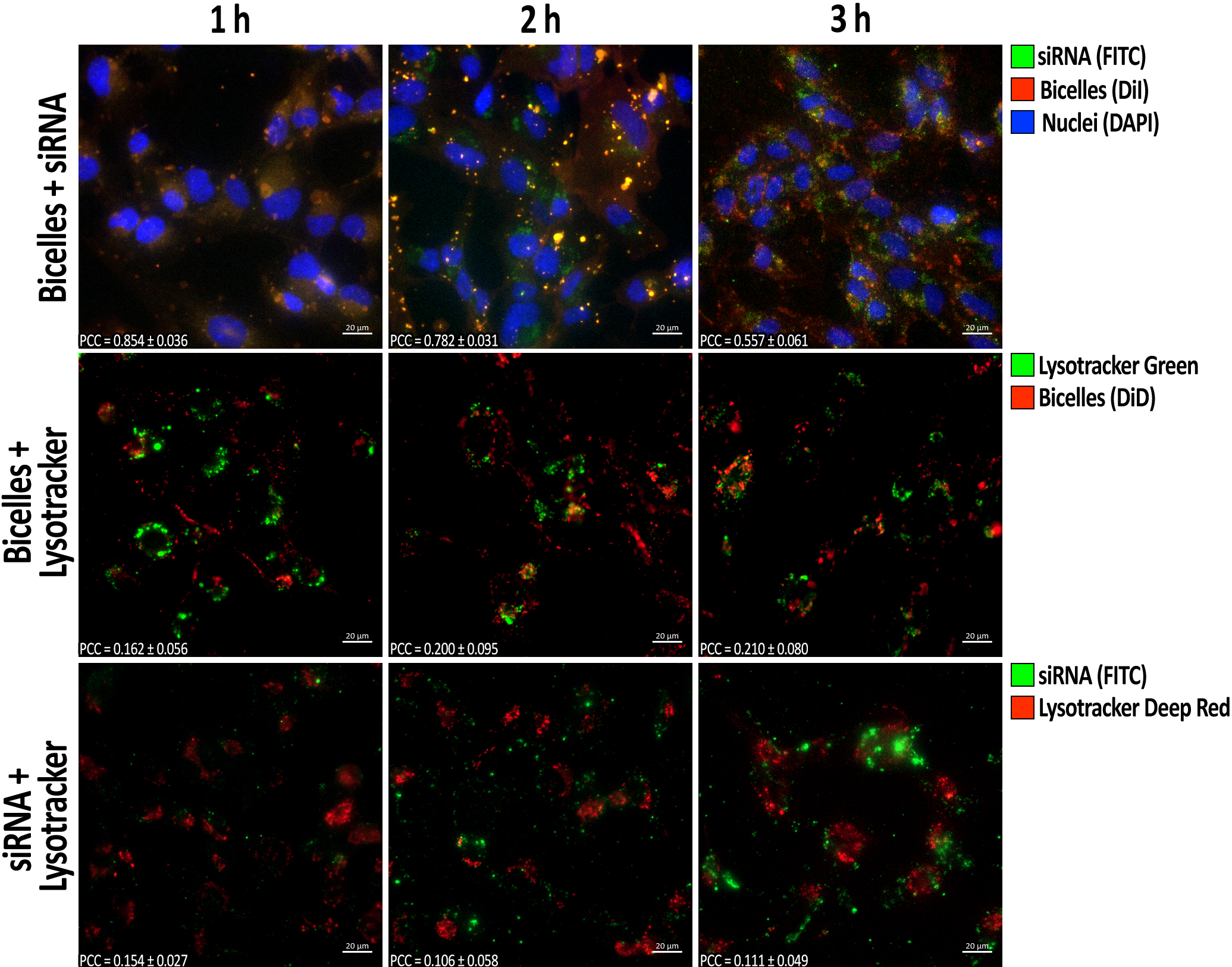 Micrographs of polarized hCMEC/D3 monolayers after treatment with siRNA-bicelle complexes at an N/P ratio of 4:1. Nuclei were stained with DAPI (blue), bicelles were laced with DiI or DiD (red), siRNA was labeled with FITC (green), and lysosomes were tagged with Lysotracker Green/Deep Red (green/red). The scale bar is 20 μm. Pearson correlation coefficients (PCC) were calculated between bicelles and siRNA, bicelles and Lysotracker, or siRNA and Lysotracker. Mean ± SD (n ≥ 6).
 Pearson correlation coefficients (PCC) between bicelles and early endosomes (Rab5), bicelles and late endosomes (Rab7), bicelles and recycling endosomes (Rab11), and siRNA and early endosomes (Rab5) at different time points (n = 12). Values above the red dotted line indicate significant colocalization.